Pulmonary embolism - ECG case study
 Written by Judy Haluka
Changes: Fix formatting of the rhythm strip
This 31-year-old male presents with sharp chest pain that started 1 hour ago. He is moderately short of breath with a pulse oximetry reading of 91% on room air. His BP is 168/98 with a pulse of 101. He appears anxious and uncomfortable. PMHX:
Unremarkable He is on no medications. His ECG is as follows:
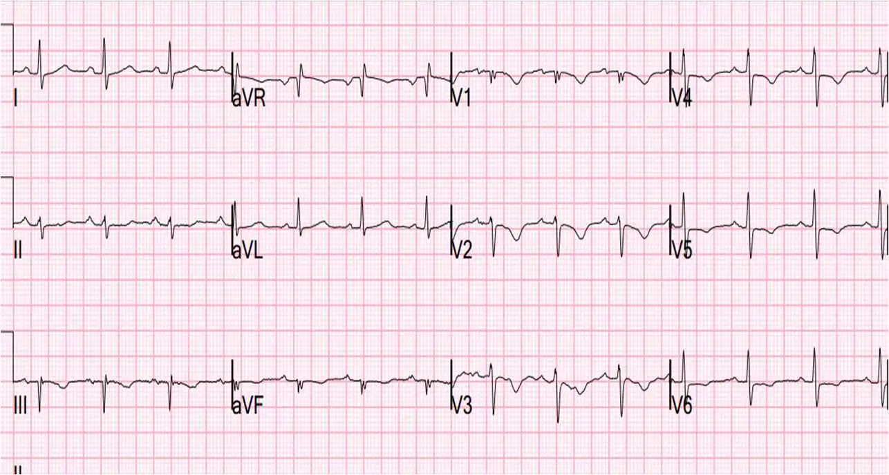
There are both precordial T-wave inversions AND T-wave inversion in lead III. These two things together are highly suggestive of pulmonary embolism. Many would assume that this represents ischemia and that he is experiencing angina. However, this ECG coupled with his history make PE much more likely.
Kosuge et al. showed that, when T-waves are inverted in precordial leads, if they are also inverted in lead III and V1, then pulmonary embolism is far more likely than ACS. In this study:
“Negative T waves in leads III and V1 were observed in only 1% of patients with ACS compared with 88% of patients with APE (p less than 0.001). The sensitivity, specificity, positive predictive value, and negative predictive value of this finding for the diagnosis of PE were 88%, 99%, 97%, and 95%, respectively. In conclusion, the presence of negative T waves in both leads III and V1 allows PE to be differentiated simply but accurately from ACS in patients with negative T waves in the precordial leads.”
Works cited
Kosuge, M., Kimura, K., Ishikawa, T, et al. Electrocardiographic differentiation between acute pulmonary embolism and acute coronary syndromes on the basis of negative T waves, Am J Cardiol. 2007; 99(6), 817-821.
Test your knowledge
Stay on track!
Would you like a reminder when your ACLS certification expires, plus study tips?
How we reviewed this article
Our experts continually monitor the medical science space, and we update our articles when new information becomes available.
- Current versionMail the author of this pageEmail
- Jan 14, 2023
Copy edited by:
Copy editorsChanges: Fix formatting of the rhythm strip- Nov 29, 2021
Reviewed by:
 Caitlin Goodwin DNP, CNM, RN Caitlin Goodwin, DNP, RN, CNM, is a Board Certified Nurse-Midwife, Registered Nurse, and freelance writer. She has over twelve years of experience in nursing practice.
- Dec 29, 2015
Written by:
Judy Haluka Judy has helped write or review several medical publications for us. Everything that she works on will clearly include Judy’s name.